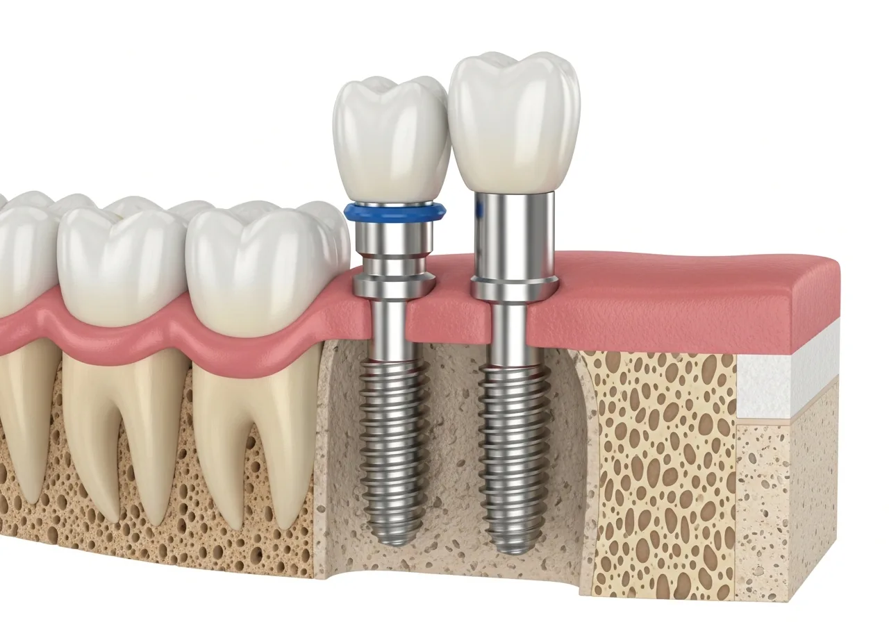

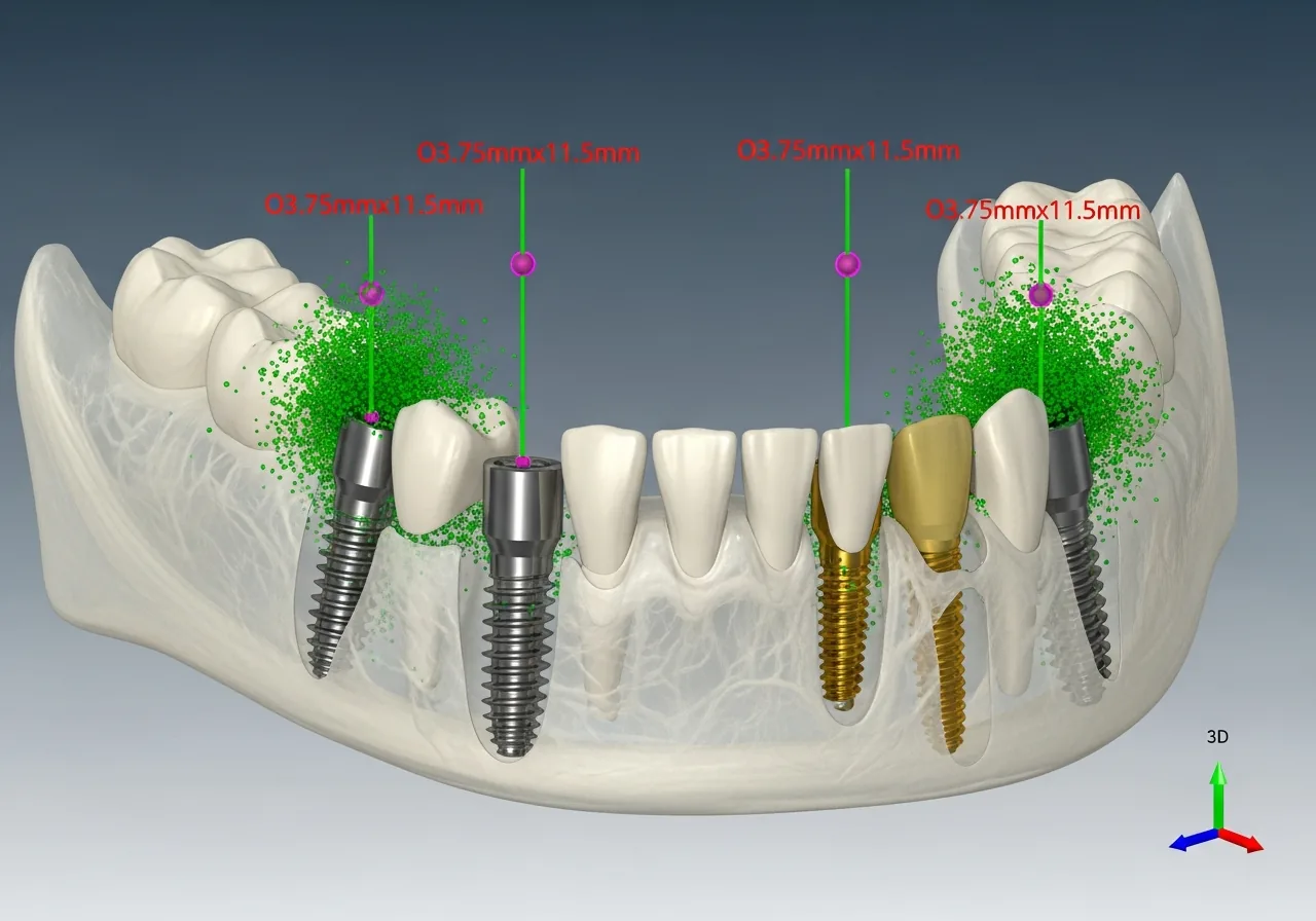
Three-dimensional implant planning on screen

3D Implant Planning as a Modern Standard

Digital volume tomography (CBCT) is, for us, the modern basis of every implant plan. It has become a firmly established part of modern implantology and oral, maxillofacial and facial surgery.

Our digital volume tomograph enables three-dimensional images of the bony facial skull. This technology opens up reliable diagnostic and therapeutic options in surgery, prosthetics, orthodontics and conservative dentistry.

Classic, two-dimensional X-ray diagnostics is often not sufficient to safely plan an implant treatment - for example, in cases of limited bone volume or a nerve close to the tooth roots. With crisp, sharp 3D views, we display the bony structures of the upper and lower jaw in detail.

What 3D planning means for you

Even before the procedure, we know exactly what to expect.

3D planning with exact measurements

Precise advance planning

The position, size and type of implant are determined even before the procedure - the implantation can be simulated in advance on screen.
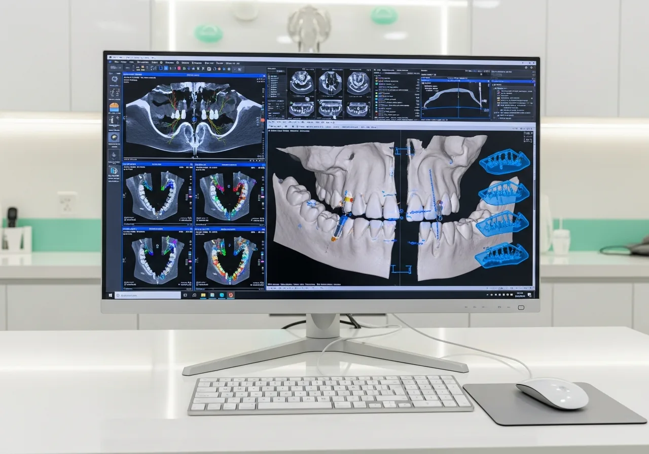
Implants in the jawbone, sensitive structures visible

Sensitive structures made visible

Nerves and other sensitive structures in the jaw are reliably identified and taken into account during planning.
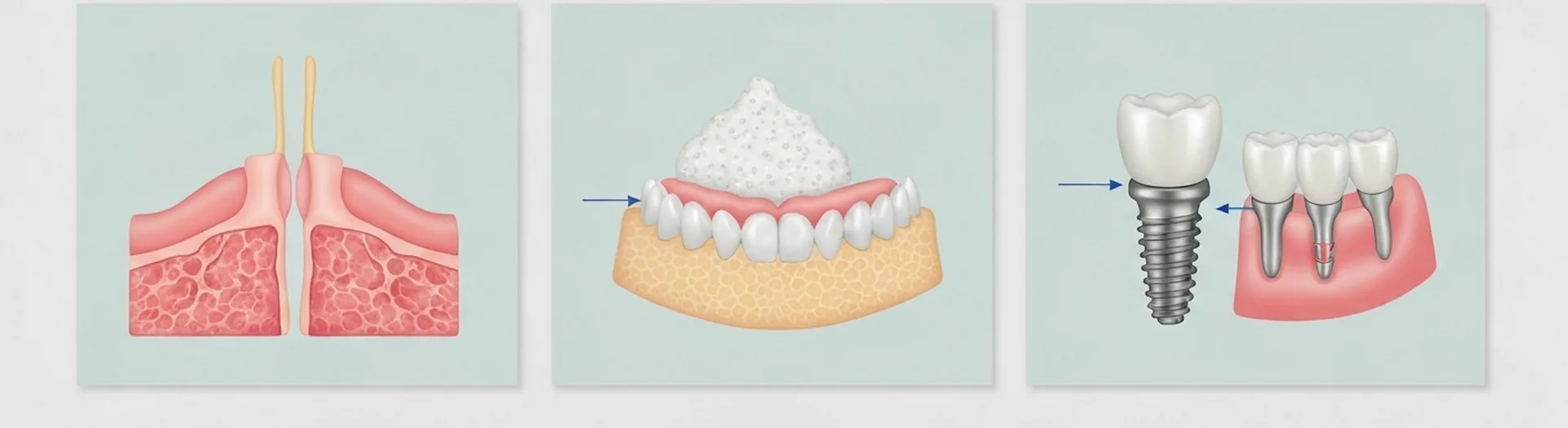
Step-by-step illustration of a possible bone graft

Less bone grafting needed

Because existing bone can be used more efficiently, an elaborate bone graft can sometimes be avoided.

X-ray monitor with low radiation exposure

Lower radiation exposure

Compared to classic computed tomography, the radiation exposure of CBCT is significantly reduced.

The Most Important Facts at a Glance

  • Digital volume tomography (CBCT) provides three-dimensional images of the jaw for precise implant planning.
  • Compared to classic computed tomography, the radiation exposure of CBCT is significantly lower.
  • With 3D diagnostics, the position, size and type of implant are determined before the procedure.
  • Sensitive structures such as nerves are made visible and protected before implantation.
  • Precise planning can often avoid the need for an additional bone graft.

Frequently Asked Questions

What is digital volume tomography (CBCT)?
CBCT is an imaging method that creates three-dimensional X-ray images of the jaw. It provides significantly more detailed information than a classic two-dimensional X-ray and is mainly used for implant planning.
Is a CBCT scan associated with more radiation than normal X-ray?
The radiation exposure of a CBCT is significantly lower than that of a classic computed tomography scan, but somewhat higher than a single two-dimensional X-ray. We use CBCT specifically where it offers real diagnostic added value.
When is 3D diagnostics necessary before implantation?
Three-dimensional imaging is particularly useful when bone volume is limited, when nerves are close to the planned implant position, or in more complex anatomical situations, in order to plan the implantation safely.